
Main menu

  • Home
  • Content
    • Current Issue
    • Ahead of Print
    • Past Issues
    • Supplements
    • Article Type
  • Specialty
    • Articles by Specialty
  • CME/MOC
    • Articles
    • Calendar
  • Info For
    • Manuscript Submission
    • Authors & Reviewers
    • Subscriptions
    • About CCJM
    • Contact Us
    • Media Kit
  • Conversations with Leaders
  • Conference Coverage
    • Kidney Week 2024
    • CHEST 2024
    • ACR Convergence 2023
    • Kidney Week 2023
    • ObesityWeek 2023
    • IDWeek 2023
    • CHEST 2023
    • MDS 2023
    • IAS 2023
    • ACP 2023
    • AAN 2023
    • ACC / WCC 2023
    • AAAAI Meeting 2023
    • ACR Convergence 2022
    • Kidney Week 2022
    • AIDS 2022
  • Other Publications
    • www.clevelandclinic.org

User menu

  • Register
  • Log in

Search

  • Advanced search
Cleveland Clinic Journal of Medicine
  • Other Publications
    • www.clevelandclinic.org
  • Register
  • Log in
Cleveland Clinic Journal of Medicine

Advanced Search

  • Home
  • Content
    • Current Issue
    • Ahead of Print
    • Past Issues
    • Supplements
    • Article Type
  • Specialty
    • Articles by Specialty
  • CME/MOC
    • Articles
    • Calendar
  • Info For
    • Manuscript Submission
    • Authors & Reviewers
    • Subscriptions
    • About CCJM
    • Contact Us
    • Media Kit
  • Conversations with Leaders
  • Conference Coverage
    • Kidney Week 2024
    • CHEST 2024
    • ACR Convergence 2023
    • Kidney Week 2023
    • ObesityWeek 2023
    • IDWeek 2023
    • CHEST 2023
    • MDS 2023
    • IAS 2023
    • ACP 2023
    • AAN 2023
    • ACC / WCC 2023
    • AAAAI Meeting 2023
    • ACR Convergence 2022
    • Kidney Week 2022
    • AIDS 2022
Review

Benign paroxysmal positional vertigo: Effective diagnosis and treatment

Shayna R. Cole, AuD, CCC-A and Julie A. Honaker, PhD, CCC-A
Cleveland Clinic Journal of Medicine November 2022, 89 (11) 653-662; DOI: https://doi.org/10.3949/ccjm.89a.21057
Shayna R. Cole
Vestibular Clinical Audiologist, The Balance Center, Fort Wayne, IN
  • Find this author on Google Scholar
  • Find this author on PubMed
  • Search for this author on this site
Julie A. Honaker
Section Head, Audiology, Head and Neck Institute, Cleveland Clinic; Adjunct Assistant Professor, Cleveland Clinic Lerner College of Medicine of Case Western Reserve University, Cleveland, OH
  • Find this author on Google Scholar
  • Find this author on PubMed
  • Search for this author on this site
  • For correspondence: [email protected]
  • Find this author on Cleveland Clinic
  • Article
  • Figures & Data
  • Info & Metrics
  • PDF
Loading

Article Figures & Data

Figures

  • Tables
  • Figure 1
    • Download figure
    • Open in new tab
    • Download powerpoint
    Figure 1

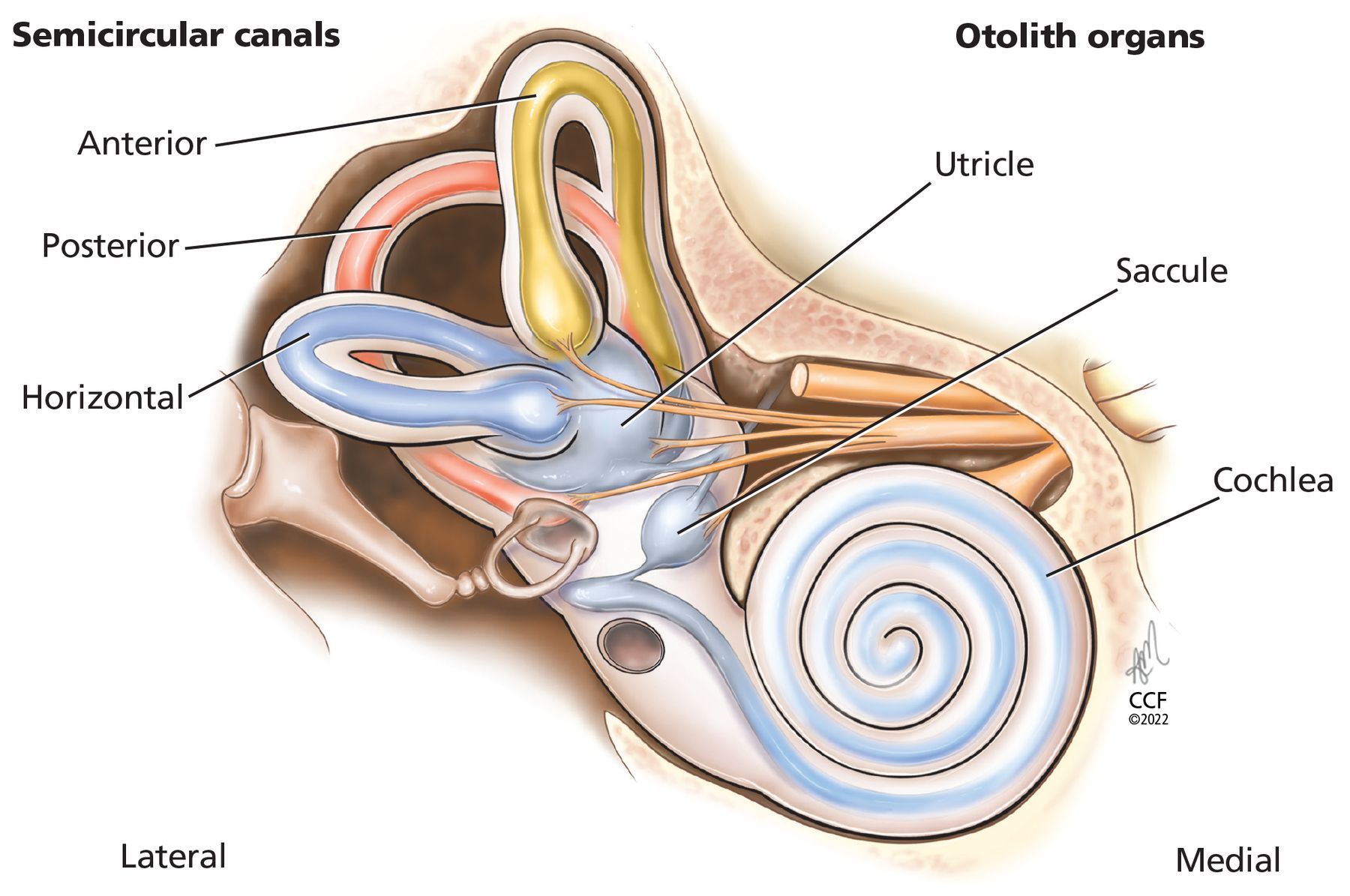
    The semicircular canals (anterior, posterior, horizontal) sense angular acceleration, and the otolith organs (saccule, uticle) sense linear acceleration, providing internal cues for orientation of position in space, movement, gaze stabilization, and postural control.

  • Figure 2
    • Download figure
    • Open in new tab
    • Download powerpoint
    Figure 2

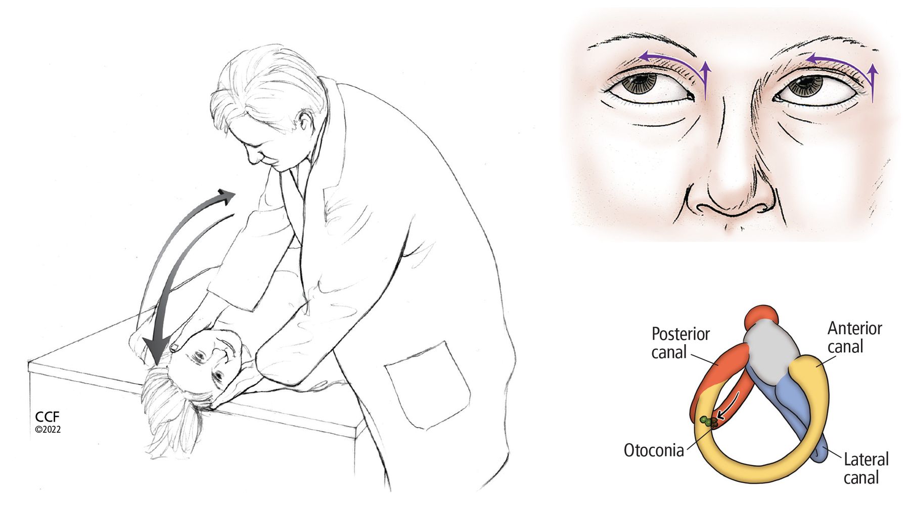
    The Dix-Hallpike maneuver to detect otoconia in the posterior or anterior semicircular canals. If the otoconia are suspected to be in the right ear, the patient sits upright with the head turned 45° to the right; if the otoconia are suspected to be in the left ear, the patient turns the head to the left. The clinician then quickly moves the patient into a head-hanging supine position and checks for signs of nystagmus, and the patient reports any symptoms (eg, dizziness, vertigo). After 60 seconds, the patient is returned to a seated position with the head still turned, and the clinician again observes symptoms and signs. During the maneuver, movement of otoconia within the right posterior semicircular canal (in the lower-right image) causes an excitatory response—ie, nystagmus—to the right and up, as the arrows indicate in the upper-right image.

  • Figure 3
    • Download figure
    • Open in new tab
    • Download powerpoint
    Figure 3

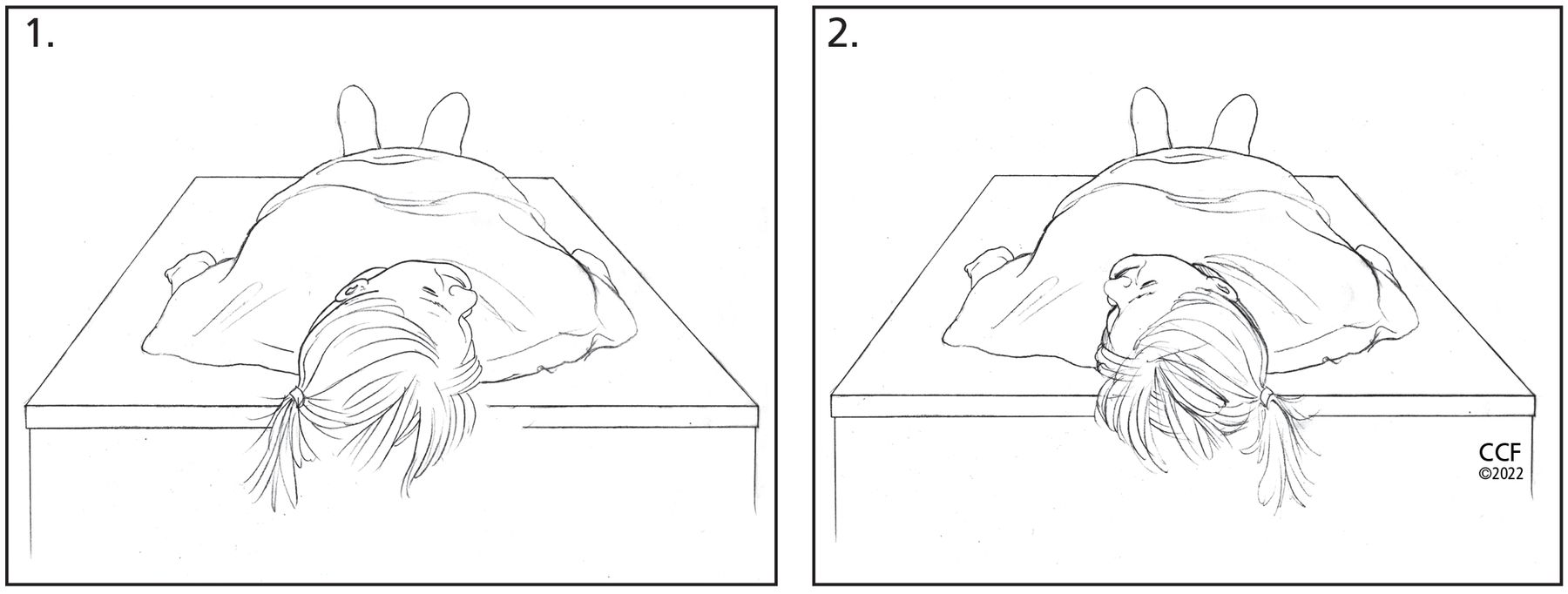
    The supine roll test to detect otoconia in the horizontal semicircular canals. (A) With the patient in a supine position, the clinician quickly rotates the patient’s head to the right and assesses for horizontal nystagmus and patient symptoms. (B) After 30 to 60 seconds, the clinician quickly rotates the patient’s head to the left and again observes for horizontal nystagmus and symptoms. The direction of nystagmus (ie, geotropic vs apogeotropic) with the head-movement changes indicates the involved horizontal canal (Table 2).

  • Figure 4
    • Download figure
    • Open in new tab
    • Download powerpoint
    Figure 4

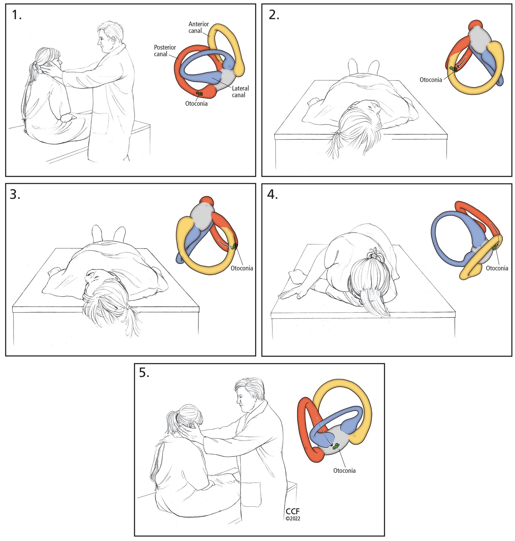
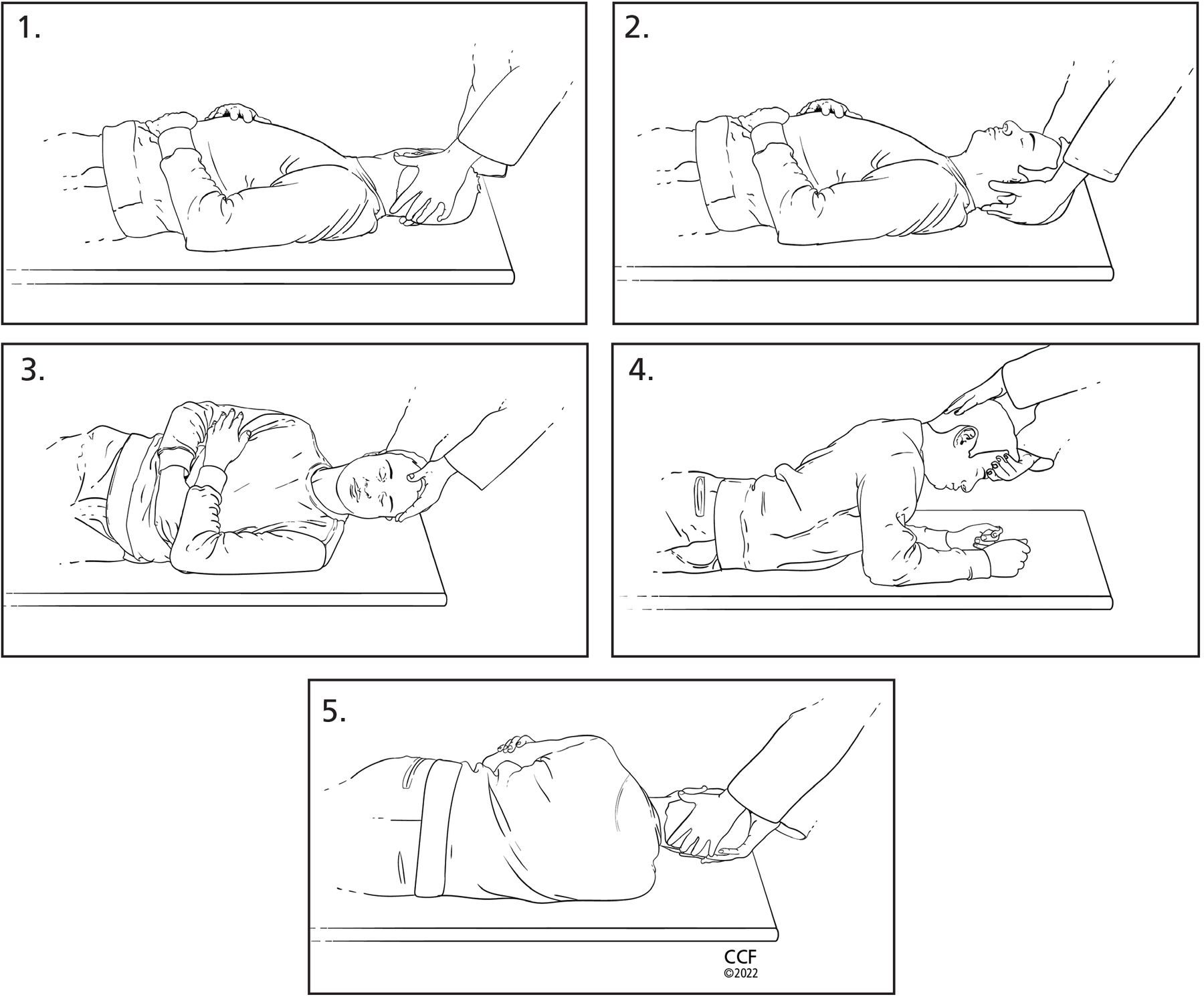
    The Epley maneuver to clear otoconia from the posterior or anterior semicircular canals:

    1. Place the patient in a seated position on the bed and turn their head 45° toward the ear with the suspected otoconia. The color insets show movement of otoconia.

    2. Quickly move the patient to a supine position with head turned and extended downward.

    3. Move the patient’s head to the other side, being careful to keep it in the correct plane. The final position after turn should be 45° toward the unaffected ear, extended downward.

    4. Assist the patient onto the unaffected side with the patient’s chin remaining 45° toward the unaffected ear (patient will be looking toward the ground in this position).

    5. Finally, help the patient back to a seated position, keeping their head turned over their shoulder.

  • Figure 5
    • Download figure
    • Open in new tab
    • Download powerpoint
    Figure 5

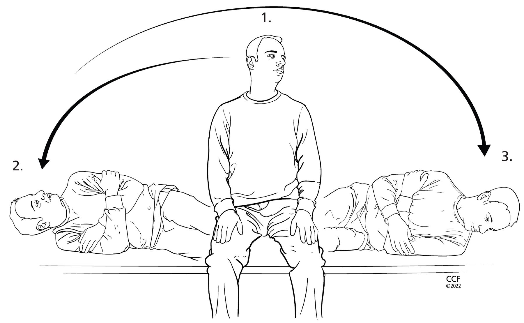
    Semont maneuver, an alternative way to clear otoconia from the posterior or anterior canals:

    1. Place the patient in a seated position on the bed and turn the head 45° away from the ear with the suspected otoconia in the vertical semicircular canal.

    2. Quickly move the patient on their side with the nose facing the ceiling.

    3. Quickly move the patient back up and onto their other side with the head in the same 45° angle with the nose facing toward the ground. The examiner then assists the patient to a seated position, keeping the head at 45° away from the ear with suspected vertical semicircular canal benign paroxysmal positional vertigo (BPPV).

    Note: The head position described in step 1 is used for posterior semicircular canal BPPV. The patient should turn the head 45° toward the ear with suspected vertical semicircular canal BPPV if the anterior semicircular canal is affected.

  • Figure 6
    • Download figure
    • Open in new tab
    • Download powerpoint
    Figure 6

    Log roll (360°) maneuver to clear otoconia from the horizontal semicircular canal:

    1. Place the patient in the supine position and turn their head 90° toward the ear with the suspected otoconia in the horizontal semicircular canal.

    2. Next, turn the patient’s head back to center, with the head elevated 30°.

    3. Maneuver the patient onto their side (90°) toward the unaffected ear.

    4. Move the patient into the prone position with elbows flexed. Note: Sometimes treatment can end in this position (called 270° maneuver).

    5. Finally, help the patient back onto their back toward the ear involved, completing a complete 360° rotation.

Tables

  • Figures
    • View popup
    TABLE 1

    Symptoms and temporal pattern of common disorders of dizziness

    DisorderSymptomsTemporal pattern
    Benign paroxysmal positional vertigo (BPPV)Head or body movement-provoked vertigoEpisodic; seconds to minutes; can have delayed latency in symptoms or fatigue of symptoms upon repeat movement
    Cervical vertigoDizziness, imbalance or lightheadedness with neck pain or changes in neck positionEpisodic; minutes to hours
    Menière diseaseVertigo with fluctuating hearing loss, aural fullness, and tinnitusSpontaneous onset; episodic; 20 minutes to 24 hours
    Vestibular neuritisVertigo without auditory symptoms, followed by head movement-provoked symptomsSudden onset; 1–3 days
    LabyrinthitisVertigo with auditory symptoms, followed by head movement-provoked symptomsSudden onset; several days
    Acoustic neuroma or vestibular schwannomaImbalance with brief episodes of dizziness; auditory symptoms; occasional neurologic symptomsGradual onset; progressive and continuous
    Superior canal dehiscenceAutophony, disequilibrium, positional vertigo, and pressure- or sound-induced symptoms of vertigoEpisodic; seconds to minutes
    Perilymphatic fistulaPressure- or sound-induced symptoms of vertigo, imbalanceSudden onset; episodic; seconds to minutes
    Vascular event (anterioinferior cerebellar artery or posterioinferior cerebellar artery stroke)Vestibular crisis event: vertigo with associated hearing loss and other neurologic symptoms and signs followed by head movement-provoked symptomsSudden onset; lasting 1–3 days
    • Based on information in references 3 and 4.

    • View popup
    TABLE 2

    Observed nystagmus patterns based on semicircular canal involvement

    Provocative maneuverNystagmus directionAffected canal
    Dix-HallpikeTo the affected side and upPosterior
    Dix-HallpikeTo the affected side and downAnterior
    Supine roll, right ear down
    Supine roll, left ear down
    Right (geotropic)
    Left (geotropic)
    Horizontal. The head-turn direction eliciting stronger nystagmus indicates the affected horizontal canal
    Supine roll, right ear down
    Supine roll, left ear down
    Left (apogeotropic)
    Right (apogeotropic)
    Horizontal. The head-turn direction eliciting weaker nystagmus indicates the affected horizontal canal
    • View popup
    TABLE 3

    Treatment maneuvers for BPPV, based on location of otoconia

    LocationTreatment maneuver
    Posterior canalEpley or Semont
    Anterior canalEpley or Semont
    Horizontal canal (geotropic-type nystagmus pattern)Log roll (360°)
PreviousNext
Back to top

In this issue

Cleveland Clinic Journal of Medicine: 89 (11)
Cleveland Clinic Journal of Medicine
Vol. 89, Issue 11
1 Nov 2022
  • Table of Contents
  • Table of Contents (PDF)
  • Index by author
  • Complete Issue (PDF)
Print
Download PDF
Article Alerts
Sign In to Email Alerts with your Email Address
Email Article

Thank you for your interest in spreading the word on Cleveland Clinic Journal of Medicine.

NOTE: We only request your email address so that the person you are recommending the page to knows that you wanted them to see it, and that it is not junk mail. We do not capture any email address.

Enter multiple addresses on separate lines or separate them with commas.
Benign paroxysmal positional vertigo: Effective diagnosis and treatment
(Your Name) has sent you a message from Cleveland Clinic Journal of Medicine
(Your Name) thought you would like to see the Cleveland Clinic Journal of Medicine web site.
CAPTCHA
Please verify that you are a real person.
Citation Tools
Benign paroxysmal positional vertigo: Effective diagnosis and treatment
Shayna R. Cole, Julie A. Honaker
Cleveland Clinic Journal of Medicine Nov 2022, 89 (11) 653-662; DOI: 10.3949/ccjm.89a.21057

Citation Manager Formats

  • BibTeX
  • Bookends
  • EasyBib
  • EndNote (tagged)
  • EndNote 8 (xml)
  • Medlars
  • Mendeley
  • Papers
  • RefWorks Tagged
  • Ref Manager
  • RIS
  • Zotero
Share
Benign paroxysmal positional vertigo: Effective diagnosis and treatment
Shayna R. Cole, Julie A. Honaker
Cleveland Clinic Journal of Medicine Nov 2022, 89 (11) 653-662; DOI: 10.3949/ccjm.89a.21057
Twitter logo Facebook logo Mendeley logo
  • Tweet Widget

Jump to section

  • Article
    • ABSTRACT
    • DIZZINESS VS VERTIGO
    • A COMMON CAUSE OF DIZZINESS AND VERTIGO
    • BRIEF EPISODES OF VERTIGO, ASSOCIATED WITH MOVEMENT
    • CAUSES OF BPPV
    • DIAGNOSTIC MANEUVERS
    • TREATMENT MANEUVERS
    • BPPV CARE PATH
    • DISCLOSURES
    • REFERENCES
  • Figures & Data
  • Info & Metrics
  • PDF

Related Articles

  • No related articles found.
  • PubMed
  • Google Scholar

Cited By...

  • A Meta-Analysis of the Epley Maneuvers Effect on Dizziness Handicap Index (DHI) Scores in Patients with Posterior Canal Benign Paroxysmal Positional Vertigo (BPPV)
  • Google Scholar

More in this TOC Section

  • High-output heart failure from arteriovenous dialysis access: A structured approach to diagnosis and management
  • Direct oral anticoagulants: Challenging prescribing scenarios in everyday practice
  • IgA nephropathy: Update on pathogenesis and treatment
Show more Review

Similar Articles

Subjects

  • Neurology

Navigate

  • Current Issue
  • Past Issues
  • Supplements
  • Article Type
  • Specialty
  • CME/MOC Articles
  • CME/MOC Calendar
  • Media Kit

Authors & Reviewers

  • Manuscript Submission
  • Authors & Reviewers
  • Subscriptions
  • About CCJM
  • Contact Us
  • Cleveland Clinic Center for Continuing Education
  • Consult QD

Share your suggestions!

Copyright © 2025 The Cleveland Clinic Foundation. All rights reserved. The information provided is for educational purposes only. Use of this website is subject to the website terms of use and privacy policy. 

Powered by HighWire