
Main menu

  • Home
  • Content
    • Current Issue
    • Ahead of Print
    • Past Issues
    • Supplements
    • Article Type
  • Specialty
    • Articles by Specialty
  • CME/MOC
    • Articles
    • Calendar
  • Info For
    • Manuscript Submission
    • Authors & Reviewers
    • Subscriptions
    • About CCJM
    • Contact Us
    • Media Kit
  • Conversations with Leaders
  • Conference Coverage
    • Kidney Week 2024
    • CHEST 2024
    • ACR Convergence 2023
    • Kidney Week 2023
    • ObesityWeek 2023
    • IDWeek 2023
    • CHEST 2023
    • MDS 2023
    • IAS 2023
    • ACP 2023
    • AAN 2023
    • ACC / WCC 2023
    • AAAAI Meeting 2023
    • ACR Convergence 2022
    • Kidney Week 2022
    • AIDS 2022
  • Other Publications
    • www.clevelandclinic.org

User menu

  • Register
  • Log in

Search

  • Advanced search
Cleveland Clinic Journal of Medicine
  • Other Publications
    • www.clevelandclinic.org
  • Register
  • Log in
Cleveland Clinic Journal of Medicine

Advanced Search

  • Home
  • Content
    • Current Issue
    • Ahead of Print
    • Past Issues
    • Supplements
    • Article Type
  • Specialty
    • Articles by Specialty
  • CME/MOC
    • Articles
    • Calendar
  • Info For
    • Manuscript Submission
    • Authors & Reviewers
    • Subscriptions
    • About CCJM
    • Contact Us
    • Media Kit
  • Conversations with Leaders
  • Conference Coverage
    • Kidney Week 2024
    • CHEST 2024
    • ACR Convergence 2023
    • Kidney Week 2023
    • ObesityWeek 2023
    • IDWeek 2023
    • CHEST 2023
    • MDS 2023
    • IAS 2023
    • ACP 2023
    • AAN 2023
    • ACC / WCC 2023
    • AAAAI Meeting 2023
    • ACR Convergence 2022
    • Kidney Week 2022
    • AIDS 2022
The Clinical Picture

Blue sclera: An overlooked finding of iron deficiency

Yasuhiro Kano, MD
Cleveland Clinic Journal of Medicine October 2022, 89 (10) 549-550; DOI: https://doi.org/10.3949/ccjm.89a.22045
Yasuhiro Kano
Fellow, Department of General Internal Medicine, Tokyo Metropolitan Tama Medical Center, Fuchū, Tokyo, Japan
  • Find this author on Google Scholar
  • Find this author on PubMed
  • Search for this author on this site
  • For correspondence: [email protected]

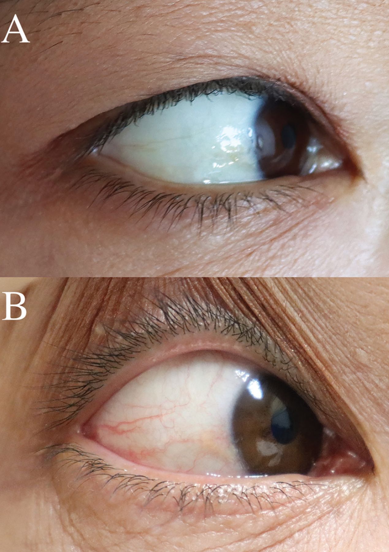
A 46-year-old woman presented with a 3-month history of progressive fatigue and shortness of breath. She had a history of uterine fibroids with heavy menstrual bleeding but had missed her gynecologic follow-up examination 2 years earlier. Physical examination revealed facial and conjunctival rim pallor, blue sclera (Figure 1A) with conjunctival rim pallor, and koilonychia. Laboratory tests were hemoglobin 4.0 g/dL (reference range 12–16), hematocrit 16.7% (37–47), mean corpuscular volume 54.8 fL (80–98), serum ferritin 0.8 ng/mL (24–307), and transferrin saturation 2.8% (20%–50%). Iron deficiency anemia was diagnosed.

Figure 1
  • Download figure
  • Open in new tab
  • Download powerpoint
Figure 1

(A) Bluish sclera with peculiar brilliancy and pale skin at presentation. (B) Normalized sclera color 3 months after iron supplementation therapy.

The patient received a blood transfusion and began taking iron supplements. Her uterine fibroids were treated with oral gonadotropin-releasing hormone antagonists. Three months later, her symptoms and physical findings, including the blue sclera (Figure 1B), had resolved, and her hemoglobin and ferritin levels had normalized.

Blue sclera is a common and useful finding of iron deficiency but is often overlooked. In 1908, Sir William Osler first described a blue discoloration of the sclera as a symptom of anemia in young girls and wrote that the eyes “have a peculiar brilliancy and the sclerotics are of a bluish color”.1 Kalra et al2 subsequently found that blue sclera is more common in patients with iron deficiency anemia (87%) than in those with other types of anemia (7%). In adult patients, blue sclera reportedly has 87% to 89% sensitivity and 64% to 94% specificity for iron deficiency anemia and iron deficiency (ie, anemia need not always be present).2,3 Blue sclera has been reported in other conditions, albeit rarely, including rheumatoid arthritis, myasthenia gravis, and long-term steroid therapy.2 Its pathogenesis is thought to involve thinning of the collagen fibers of the sclera due to iron deficiency, which allows the bluish color of the underlying uvea to become visible.

DISCLOSURES

The author reports no relevant financial relationships which, in the context of their contributions, could be perceived as a potential conflict of interest.

  • Copyright © 2022 The Cleveland Clinic Foundation. All Rights Reserved.

REFERENCES

    1. Osler W
    . Primary or essential anemia. In: The Principles and Practice of Medicine. 6th ed. New York, New York: D. Appleton and Company: 1908: 721–724.
    1. Kalra L,
    2. Hamlyn AN,
    3. Jones BJ
    . Blue sclerae: a common sign of iron deficiency? Lancet 1986; 2(8518):1267–1269. doi:10.1016/s0140-6736(86)92688-7
    1. Kotsev I,
    2. Pavlov S,
    3. Zheleva D,
    4. Gencheva T
    . Sinie sklery pri zhelezodefitsitnykh sostoianiiakh [Blue sclera in iron deficiency conditions]. Klin Med (Mosk) 1991; 69(8):85–86.

Navigate

  • Current Issue
  • Past Issues
  • Supplements
  • Article Type
  • Specialty
  • CME/MOC Articles
  • CME/MOC Calendar
  • Media Kit

Authors & Reviewers

  • Manuscript Submission
  • Authors & Reviewers
  • Subscriptions
  • About CCJM
  • Contact Us
  • Cleveland Clinic Center for Continuing Education
  • Consult QD

Share your suggestions!

Copyright © 2025 The Cleveland Clinic Foundation. All rights reserved. The information provided is for educational purposes only. Use of this website is subject to the website terms of use and privacy policy. 

Powered by HighWire